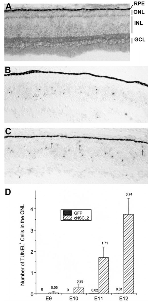
Figure 6.

Photoreceptor degeneration in retinas misexpressing cNSCL2. (A) Anti-P27 immunohistochemistry of an E9 retina infected with RCAS-cNSCL2. The ONL appeared normal. (B, C) TUNEL+ cells in E11 retinas infected with RCAS-GFP (B) or RCAS-cNSCL2 (C). TUNEL+ cells were absent in the ONL of the control (B) but were present in experimental retina (C, arrows). (D) Quantification of TUNEL+ cells in the ONL of RCAS-cNSCL2, or RCAS-GFP, retinas at different developmental stages. Shown are means ± SD of TUNEL+ cells per view area under a ×20 objective. Each data point represents three retinas with more than 20 view areas scored in each retina.
