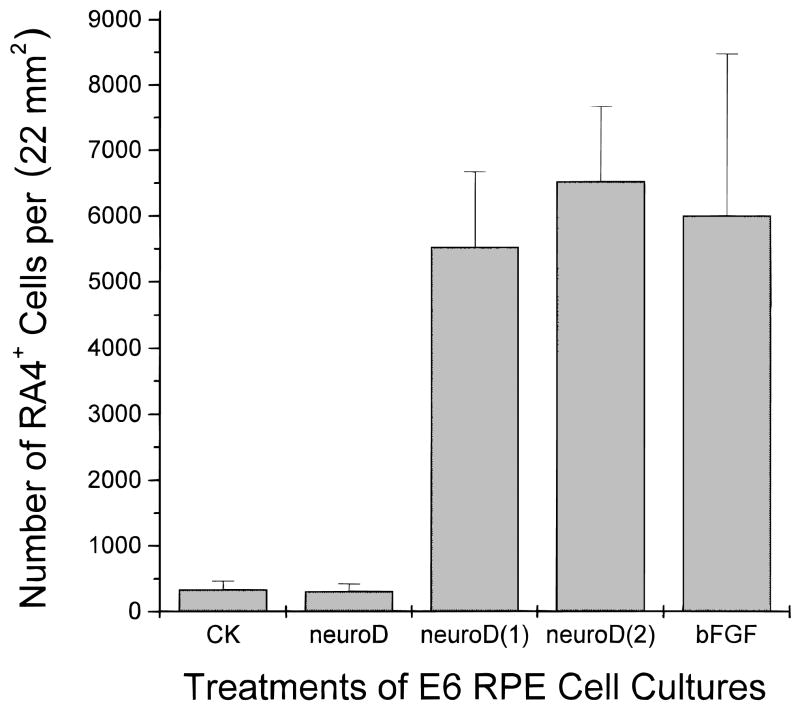
Fig. 3.
Quantification of RA4-positive cells in E6 RPE cultures under different treatments. The number of cells positive for RA4 increased dramatically when RPE cells were cultured in the presence of bFGF alone (bFGF), compared to the control (CK) or to those ectopically expressing neuroD alone (neuroD). In the presence of neuroD and bFGF, either added at the same time [neuroD(1)] or bFGF added 4 days after the addition of neuroD-expressing retrovirus [neuroD(2)], the number of RA4-positive cells was high and not statistically different from that in dishes treated with bFGF alone.

