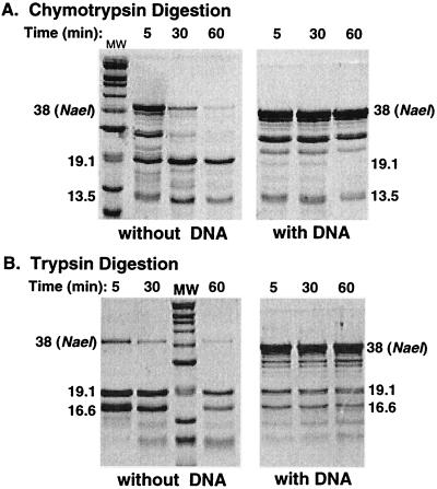
Figure 4.
Coomassie brilliant blue stained SDS-polyacrylamide gel showing pattern of polypeptide fragments produced by limited chymotrypsin (A) and trypsin (B) digestion of NaeI protein in the presence or absence of DNA. Digestion times are shown above the lanes. The Mr of protease-resistant fragments are indicated alongside the gel image and are based on the molecular weight (MW) markers described in the legend to Fig. 2.

