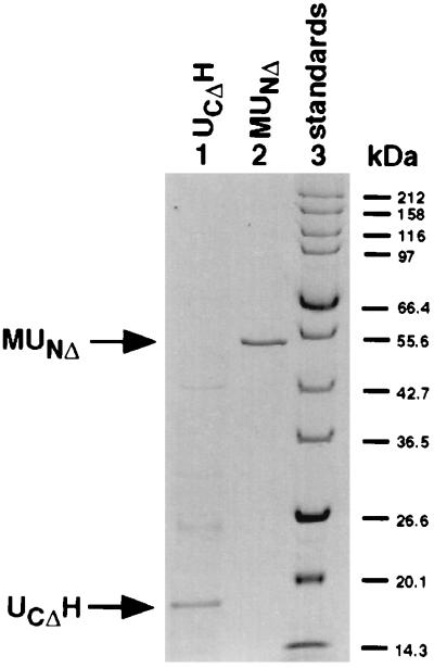
Figure 2.
Purification of the fusion proteins MUNΔ and UCΔH. MUNΔ and UCΔH were isolated as described in Materials and Methods. Approximately 5 μg UCΔH (lane 1), 0.2 μg MUNΔ (lane 2), and broad-range protein marker (New England Biolabs, lane 3) were subjected to SDS/PAGE by using the standard protocol (13). The gel was stained with Coomassie blue.

