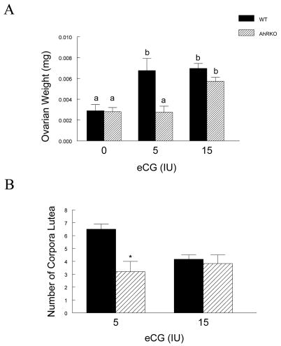
Figure 3. The Effect of Ahr Deletion on Ovarian Weight and the Number of Corpora Lutea in Response to Gonadotropin Treatment.
Panel A: AhRKO and WT mice were injected with eCG (5 IU and 15 IU) on postnatal days 25–28 and ovarian weight was measured as described in methods. Each bar represents the mean ± SEM. Bars with different letters are significantly different from each other (n = 3 – 8, p = 0.92 for 0 IU eCG in WT vs. 0 IU eCG in AhRKO; p ≤ 0.013 for 5 IU eCG in WT vs. 5 IU eCG in AhRKO; p = 0.13 for 15 IU eCG in WT vs. 15 IU eCG in AhRKO). Panel B: AhRKO and WT mice were injected with eCG (5 IU and 15 IU) on postnatal days 25–28 and then the number of corpora lutea were counted in each ovary. Each bar represents the mean ± SEM. Asterisks indicate statistically significant differences between genotypes (n = 3 – 8, p ≤ 0.02 for 5 IU eCG in WT vs. 5 IU eCG in AhRKO; p = 0.68 for 15 IU eCG in WT vs. 15 IU eCG in AhRKO).

