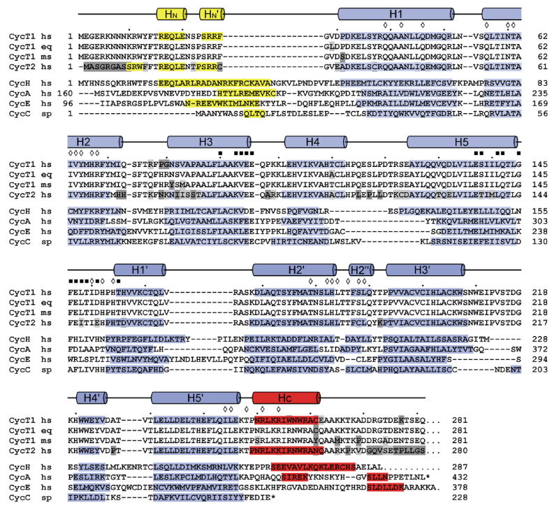
Figure 2.

Structure-based sequence alignment of the cyclin box repeats from various cyclin proteins. The alignment is based on the structures of human Cyclin H (1JKW, ref. 30), human Cyclin A (1FIN, ref. 8), human Cyclin E (1W98, ref. 12), Cyclin C from S. pombe (1ZP2, ref. 32) and human CycT1 (2PK2, this study). Secondary structure elements of CycT1 are indicated on top, helices of cyclins H, A, E and C are displayed by bars with the N- and C-terminal helices coloured yellow and red, respectively. Residues within the interface of the two repeats in CycT1 are indicated by diamonds. Residues of CycT1 that are proposed to interact with Cdk9 are marked by black squares.
