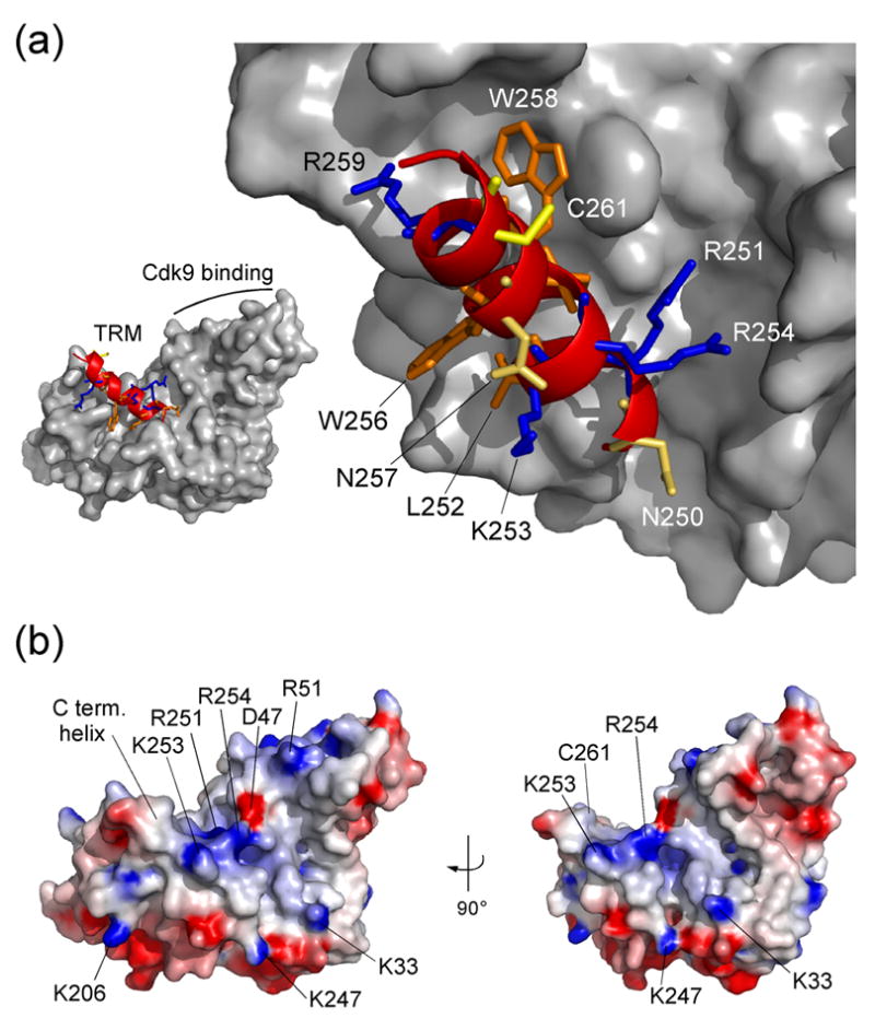
Figure 3. The Tat-TAR recognition motif in Cyclin T1.

(a) Conformation of the C-terminal region from residue 247 to 263 as ribbon plot within the CycT1 structure. Positively charged residues K253 and R254, polar asparagines N250 and N257, and the aromatic W256 are accessible on the surface of the TRM structure. In contrast, side chains of R251, L252, I255 and W258 connect the C-terminal helix to the cyclin box repeats. Cystein 261 which might form an intermolecular zinc-finger with Tat is fully exposed on the surface. (b) Electrostatic surface potential of CycT1 (displayed from −8 kBT to +8 kBT). Basic residues of the TRM form a combinatorial surface with the first cyclin box repeat in between both cyclin boxes that could attract negatively charged RNA molecules of 7SK or TAR.
