
Figure 3.
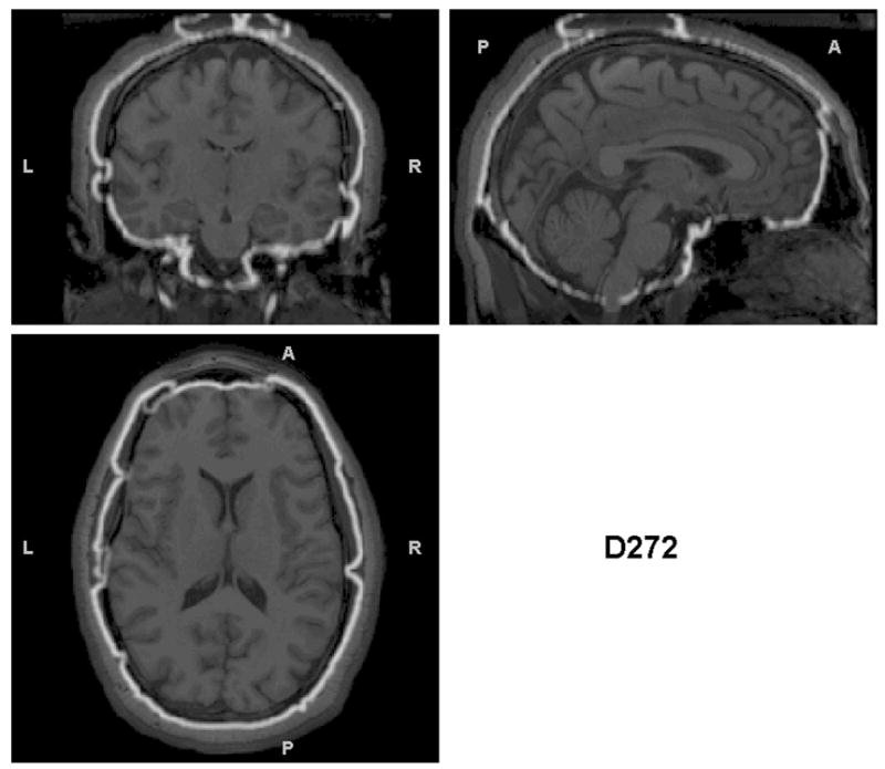
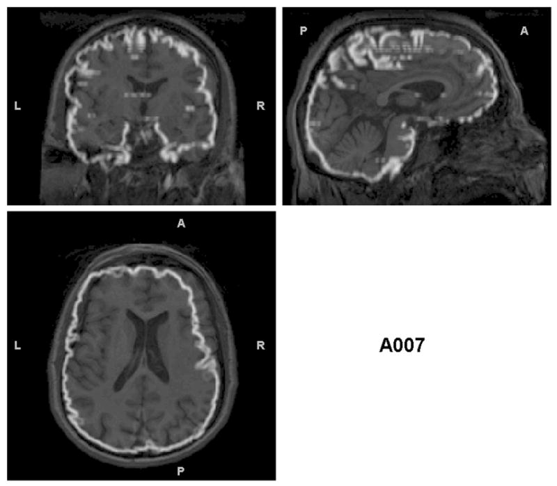
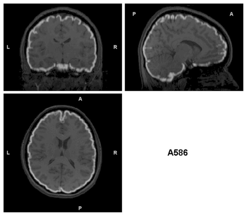
Triplanar view of the outer boundary of the GM segmentation (outlined in white) using the non-brain-extracted T1s as input for the subjects presented in Figure 2. The GM segmentation for (a) is outside the border of the brain and in many cases within the skull. The segmentation in (b) has less (but some) non-brain tissue included in the outline of the GM segmentation. However, this example shows another problem that is not obvious without examining these boundaries – that there are brain regions excluded from the GM segmentation (visible in the sagittal and coronal views in the anterior right). (c) the GM segmentation is well aligned with the brain boundary, with no problems indicated.