
Figure 5.
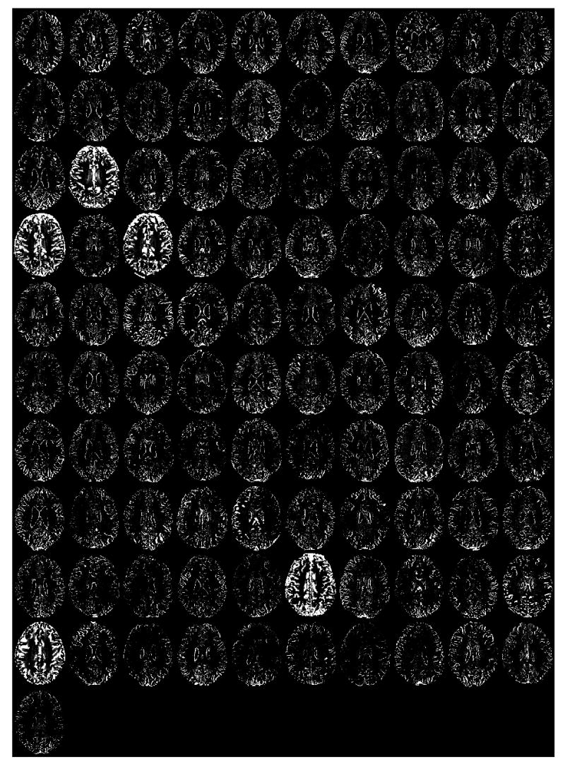
Midaxial slice (for each of the 101 research subjects) of the difference in the gray matter SPM2 segmentations computed by subtracting the segmentation using brain-extracted inputs from that using non-brain extracted inputs. Bright areas denote non-brain matter inclusions in the segmentation using non-brain extracted inputs.