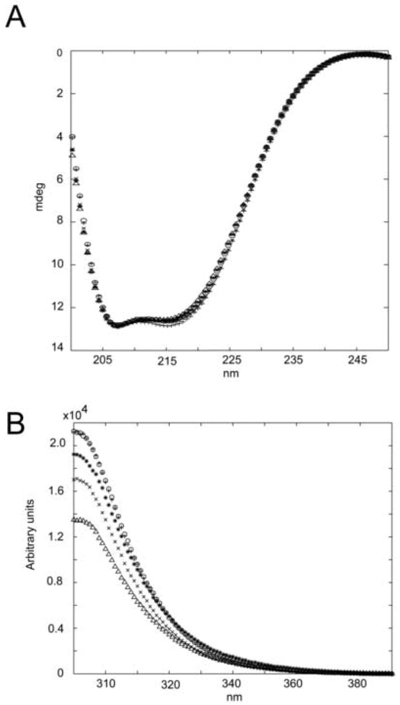
Figure 1. Circular dichroism and tyrosine emission fluorescence.

A) Far-UV circular dichroism spectra; B) tyrosine emission fluorescence of Spo0F mutants; “x” Y13A, “^” L66A, “*” I90A, “0” H101A and “+” wild type. Results confirm similar secondary structures between all mutant proteins and wild type, indicating no major conformational changes upon mutation.
