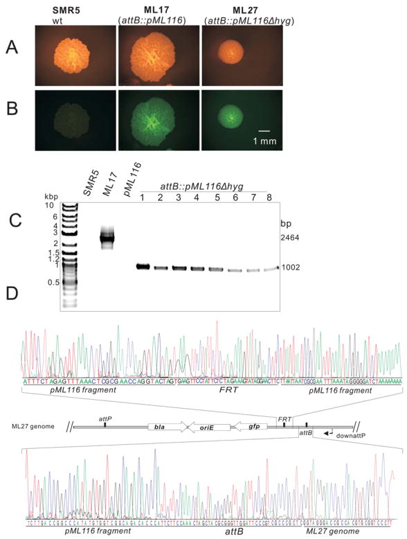
Fig. 2. Integration and removal of a FRT-hyg-FRT cassette from M. smegmatis.

A and B: Microscopy of single colonies of M. smegmatis SMR5 (wt), ML17 (attB::pML116) and ML27 (attB::pML116Δhyg). Pictures of the same colonies were taken with an Olympus SZX12 (Model U-ULH) stereomicroscope equipped with a Zeiss AxioCam MRc camera and an Olympus fluorescence illuminator (100W mercury lamp) using a 20-fold magnification. A fluorescence filter cube (excitation: 470 nm, emission: 500 nm) was used to reveal fluorescence of the colonies. Note that the sizes of the colonies are different because they were incubated for different days. The scale bar is shown.
C: Agarose gel (1%) analysis of the PCR products amplified from chromosomal DNA. The primer pair MGR19 and downattP yielded no fragments for SMR5 and pML116 and fragments of 2,464 bp and 1,002 bp for ML17 and 8 different clones picked after flpm expression.
D: Partial sequencing results of the amplified 1,002 bp fragment from clone #1 which was named M. smegmatis ML27 (lane 1 of C) using primer downattP. The locations of the FRT and attB sites and parts of the pML116 fragment in the genome of ML27 are shown in the map.
