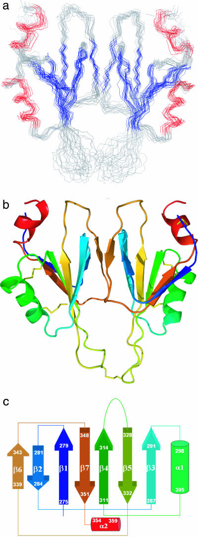
Fig. 1.
Solution structure of the A4 dimer. (a) The backbone overlay of 14 low-energy structures of FXI A4. Colored segments indicate β-sheet (blue) and helices (red). (b) Ribbon representation of the two domains of the dimer rendered in colors ranging from blue (N-terminal) to red (C-terminal). (c) Topology diagram of the NMR structure of the FXI A4 domain. The color scheme follows that in b.

