
Figure 3. Lzts1−/− MEF displayed mitotic defects.
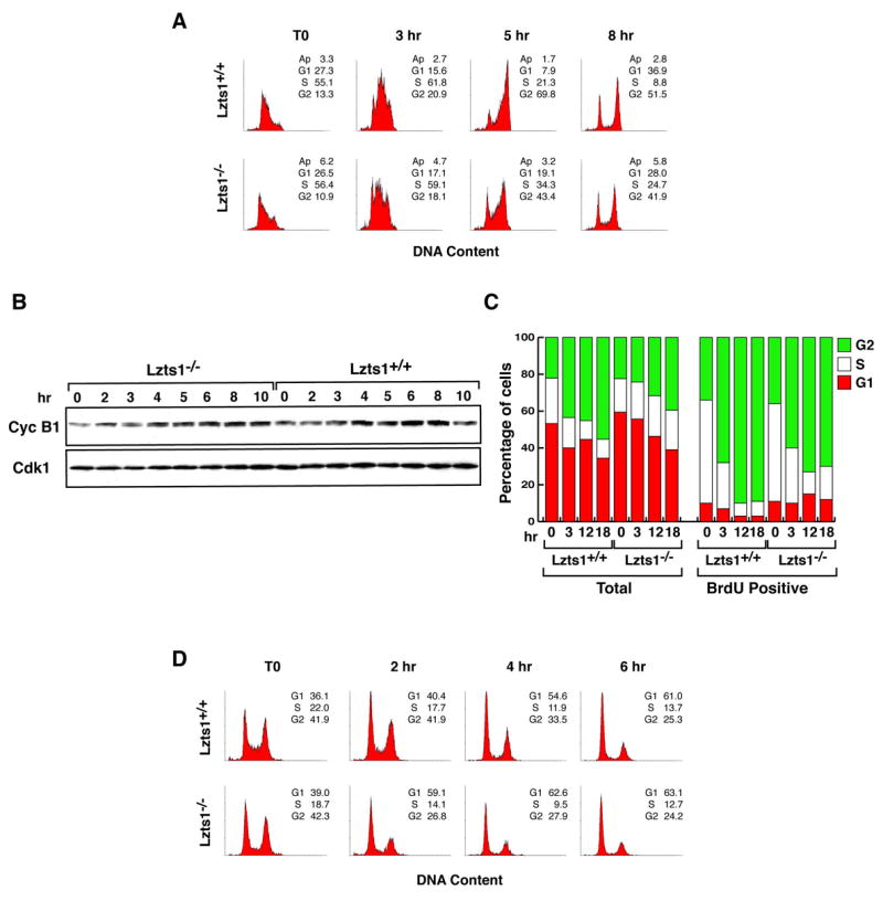
A. Flow cytometry analysis of Lzts1+/+ and −/− MEFs synchronized at the G1/S transition by double thymidine block and released for the indicated time. B. Expression of Cyclin B1 and Cdk1 proteins in cells treated as described in A. C. Evaluation of cell cycle progression in cells labeled with BrdU for 1 hour and then exposed to nocodazole for the indicated time. The percentage of total or BrdU labeled cells in each phase of the cell cycle is shown. D. Flow cytometry analysis of Lzts1+/+ and −/− MEFs treated for 10 hours with Hoechst 33342 (Left panels, T0) and then released for the indicated time.