
Figure 5. Lzts1 absence results in decreased Cdk1 activity and chromosome missegregation.
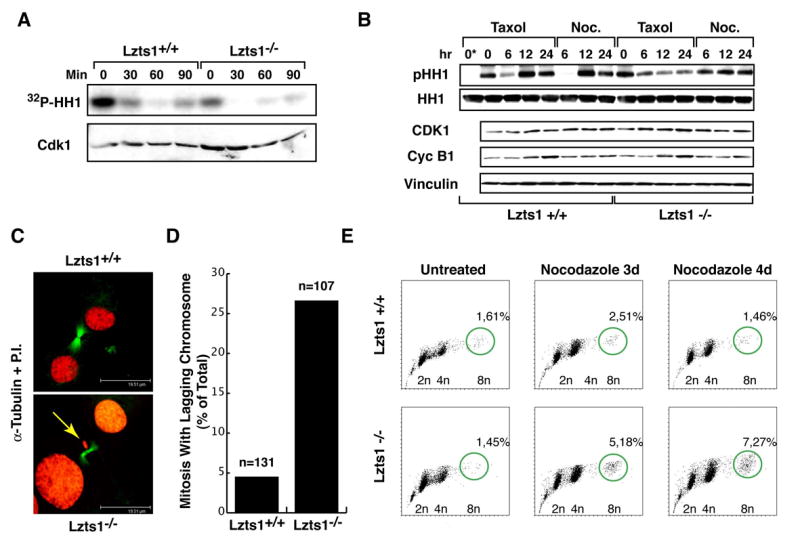
A. Cdk1 associated kinase activity in MEFs collected by mitotic shake-off (0) and released for the indicated times. In the lower panels the levels of Cdk1 are shown. B. Expression of Cyclin B1 and Cdk1 proteins and Cdk1 associated kinase activity in cells treated as indicated. Histone H1 (HH1) and Vinculin were used to normalize the kinase assay and the proteins loaded on the blot respectively. 0* lane is the negative control of kinase reaction in which the IP proteins have not been added. C. Immunofluorescence analysis of Lzts1+/+ and −/− mitotic cells stained for α-tubulin (green) and P.I. (red). A representative confocal section of a normal telophase in +/+ (upper panel) and of a telophase with missegregated (lagging) chromosomes (yellow arrows in lower panel) in −/− cells are shown. Scale bar is 19.5 μm. D. Quantification of mitosis presenting missegregated chromosomes in Lzts1+/+ and −/− MEFs examined 8 hours after double thymidine block. n is the total number of mitosis analyzed. E. Flow cytometry analysis of Lzts1+/+ and −/− MEFs treated with 1μM nocodazole, as indicated. The percentage of the 8n population, representing the mean of two independent experiments on two different MEF populations, is reported.