
Figure 6. Loss of Lzts1 impairs Cdc25C-Cdk1 interaction in mitotic cells.
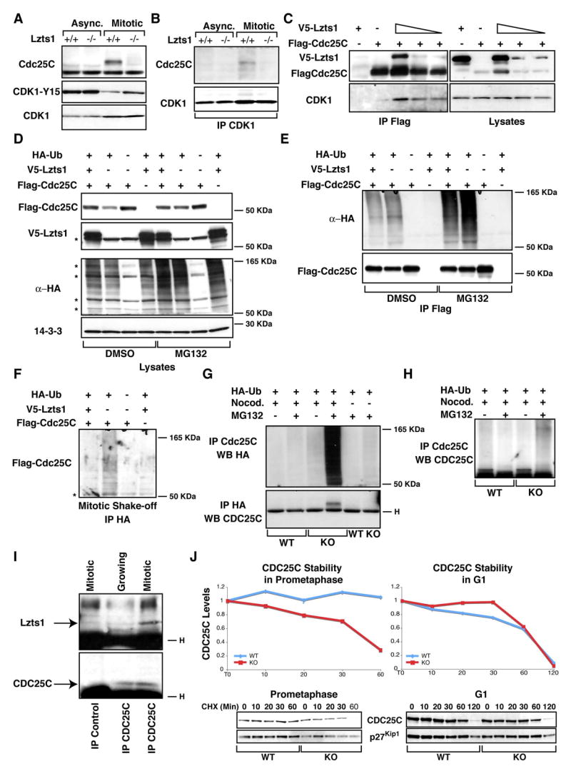
A. Western blot analysis of Cdc25C, Cdk1 and phosphoY15-Cdk1 proteins in asynchronously growing (Async.) or in mitotic (Mitotic) Lzts1+/+ and −/− MEFs. B. Western blot analysis of Cdc25C and Cdk1 in lysates from Lzts1+/+ and −/− MEFs as in A, immunoprecipitated with anti-Cdk1-Ab. C. Western blot analysis of hLzts1, hCdc25C and hCdk1 in lysates (right panels) and Flag-IP (Left panels) in 293 cells transfected with Flag-hCdc25C and increased amounts of V5-hLzts1. D. Expression of hLzts1 and hCdc25C proteins in 293 transfected with the indicated vectors and treated with nocodazole for 6 hours in the presence or not of the proteasome inhibitor MG132 (25 μM). Expression of Ha-Ubiquitin and 14-3-3 proteins was used as loading control. E. Cell lysates described in D were IP with an anti Flag-ab and analyzed for Ha-Ubiquitin and Cdc25C expression. F. 293 cells treated as in D were isolated by mitotic shake off and cell lysates were IP with an anti Flag-HA ab and analyzed for Cdc25C expression. G. Proteins from Lzts1+/+ and −/− fibroblasts transduced with HA-Ubiquitin and hCdc25C retroviruses and then isolated by mitotic shake-off were IP with either anti-Cdc25C or anti-HA Ab and probed for HA-Ubiquitin or Cdc25C expression as indicated. H. Proteins from Lzts1+/+ and −/− fibroblasts treated as in G were IP and probed with an ab specific for the Cdc25C phosphatase. * Indicates non specific bands. I. Proteins from 293T cells isolated from exponentially growing or mitotic cells were IP with an ab specific for the Cdc25C phosphatase (or control ab) and probed for Lzts1 (Upper panel) and Cdc25C (Lower panel). J. Cdc25C stability in mitotic (Left panels) or G1 cells (Right panels). The protein stability of Cdc25C was analyzed in prometaphase cells (nocodazole-arrested) and in asynchronous cells by treatment with cycloheximide for the indicated time points (min). Quantification of proteins expression was obtained scanning the blots with the Biorad GS100 scanner interfaced with the Quantity One software