
Figure 7. Regulation of Cdc25C activity by Lzts1 is necessary for the proper mitotic progression.
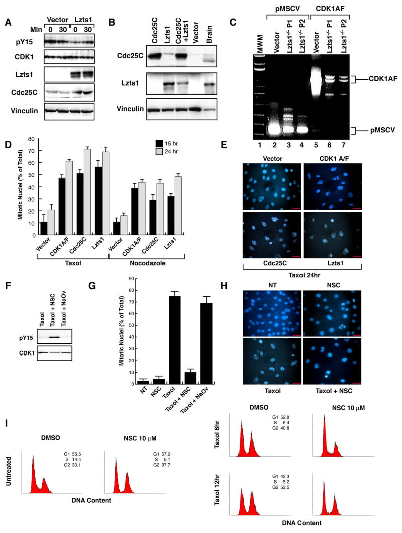
A. Western blot analysis of phosphoY15-Cdk1, Cdk1, Cdc25C, Lzts1 and Vinculin (loading control) proteins in mitotic Lzts1−/− MEFs transduced with an empty vector or with a vector encoding for the Lzts1 protein. Lysates were collected at 0 or 30 minutes after the mitotic shake-off. B. Western blot analysis of Cdc25C, Lzts1 and Vinculin (loading control) proteins in Lzts1−/− MEFs transduced with the indicated vectors. Mouse Brain lysate was used as a positive control for Lzts1 expression. C. RT-PCR analysis of Cdk1AF expression in Lzts1−/− MEFs transduced as indicated. The transduced DNA plasmids (vector) were used as positive control. D/E. Evaluation of mitotic nuclei in Lzts1−/− MEFs transduced as indicated. Typical fields of Hoechst nuclear staining are shown in E and the mean of three independent experiments is reported in D. F. Western blot analysis of Cdk1 and phosphoY15-Cdk1 expression in Lzts1+/+ MEFs treated for one hour with DMSO (NT), NSC or Na3VO4 and then with taxol for 18 hours. G/H. Evaluation of mitotic nuclei in Lzts1+/+ MEFs treated as in F. Data (mean± SD) represent the mean of a least three independent experiments performed in duplicate. I. Flow cytometry analysis of Lzts1+/+ and −/− MEFs treated with vehicle (DMSO) or NSC 10 μM for one hour and then, after the NSC wash out, exposed to 100nM Taxol for the indicated time. Scale bars 20μm.