
Fig. 5.
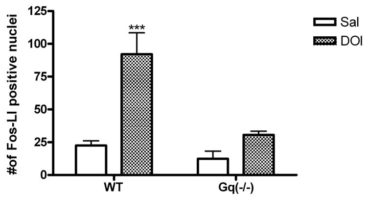
Quantitative analysis of number of FOS-Li positive nuclei in the medial prefrontal cortex of wild-type and Gαq(−/−) mice. Values are numbers of FOS-Li positive cells (mean ± S.E.M. within area of analysis; n=6). DOI-induced FOS expression in the medial prefrontal cortex is abolished in Gαq(−/−) mice. (***) p<0.001 relative to saline control group determined by a two-way ANOVA.