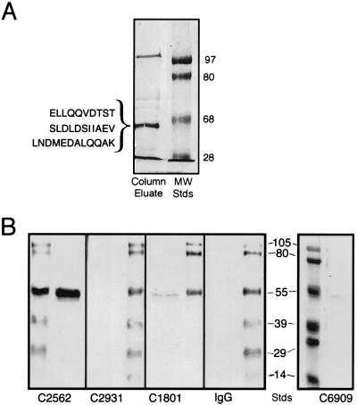
Figure 1.
(A) Purification of HK binding proteins. Biotin–HK was coupled to Ultralink Immobilized Streptavidin gel to prepare a HK affinity column as described in Experimental Procedures. HUVEC lysates were applied to the column and bound protein was eluted with 0.2 M glycine (pH 2.8) (Column Eluate). The letters to the left of the left lane represent three amino acid sequences from two separate occasions obtained from tryptic digests of the shown 54-kDa band. The lane and numbers on the right represent molecular mass standards (MW Stds) in kilodaltons. The figure is a photograph of a Coomassie blue R-250-stained 8% SDS/PAGE. (B) Immunoblot of HUVEC lysates with anti-cytokeratin antibodies. HUVEC lysates were prepared as indicated in Experimental Procedures. After electrophoresis of the lysate on 11% SDS/PAGE, the samples were transferred to nitrocellulose. Strips containing lysate were cut out and placed individually in containers containing mAbs C2562, C2931, C1801, mouse IgG, or C6909 in 0.01 M sodium phosphate and 0.15 M NaCl, (pH 7.4) containing 1% BSA and 0.01% Tween 20. After each strip was incubated and washed, the nitrocellulose was incubated with a rabbit anti-mouse antibody conjugated with horseradish peroxidase and then developed with 4-chloro-1-naphthol substrate. The antibody name is placed under the lane that characterizes the presence or absence of a band specific for cytokeratin 1. The numbers between the two photographs of the nitrocellulose membrane represent molecular mass standards in kilodaltons. The other stained lanes in each panel represent molecular mass standards.

