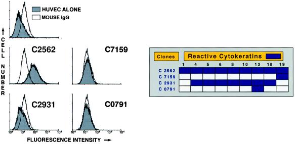
Figure 3.
Flow cytometry of HUVEC. Suspensions of washed, unfixed, and nonpermeabilized HUVEC were incubated with mouse IgG (unshaded curves) or mAbs C2562, C7159, C2931, or C0791, each added at 1/100 dilution. The binding of these antibodies on the HUVEC membrane was detected with a secondary antibody labeled with FITC. The flow cytogram of HUVEC alone not treated with any Ig is shown in the upper left. The box to the right of the flow cytograms represents the names of the antibody clones and the cytokeratins they react to. The data presented are representative of three experiments.

