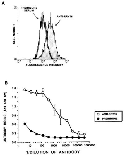
Figure 4.
(A) Flow cytometry with anti-RRY16 antisera and its preimmune serum. Washed HUVEC in suspension in Hepes–Tyrode’s buffer were incubated with 1/100 dilution of anti-RRY16 antiserum or preimmune serum for 1 h at 4°C. After centrifugation and resuspension in Hepes–Tyrode’s buffer, they were incubated with a mouse anti-goat antibody conjugated with FITC. The flow cytogram shown is a representative study of two cytograms. (B) Binding of anti-RRY16 antisera or its preimmune serum to cytokeratin. Purified cytokeratin (1 μg/well) was coupled to microtiter plate wells in 0.1 M Na2CO3 (pH 9.6) overnight at 4°C. After washing the wells with 0.01 M sodium phosphate and 0.15 M NaCl (pH 7.4), the indicated dilution of anti-RRY16 antiserum or its preimmune serum was added to the microtiter plate wells. The presence of antibody bound to the cytokeratin-coated wells was detected by using a mouse anti-goat antibody conjugated with peroxidase and peroxidase substrate. The data presented are the mean ± SEM of three determinations at each dilution.

