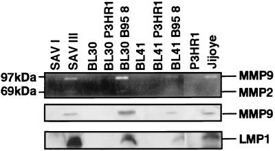
Figure 1.
MMP9 and MMP2 activity in EBV-infected type I and type III lymphoblastoid cells. SAV I is a type I BL line, and SAV III is a sister type III line. BL30 and BL 41 are EBV-negative BL lines. BL30-P3HR-1, BL30-B95–8, BL41-P3HR-1, and BL41-B95–8 lines are infected with P3HR-1 virus (EBNA2 deleted) or B95–8 virus. Jijoye is the parental BL line of P3HR-1 and has intact EBNA2 gene. (Top) Gelatinolytic activity; (Middle) Western blot of MMP9; (Bottom) Western blot of LMP1. Lymphoid cells (2.0 × 10⁶) were cultured in 1 ml media for 48 hr. After centrifugation, supernatant fluid was prepared for zymography and the pellet for Western blot analysis as described in Materials and Methods.

