
Figure 4. In vitro angiogenesis.
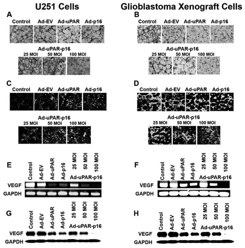
H & E staining. Endothelial cells were grown in conditioned medium, collected from U251 and glioblastoma xenograft cells infected with indicated adenoviral constructs as described in Materials & Methods, for 72 h. Then, the cells were stained with hematoxylin and eosin and capillary network formation was observed under a laser scanning confocal microscope. (A) Endothelial cell network formation in the presence of conditioned medium collected from indicated recombinant virus-treated U251 cells. (B) Endothelial cell network formation in the presence of conditioned medium collected from indicated recombinant virus-treated glioblastoma xenograft cells. Factor VIII staining. U251 and glioblastoma xenograft cells, seeded in eight-well chamber slides, were incubated overnight and infected with the indicated MOI of Ad-EV, Ad-uPAR, Ad-uPAR-p16 or Ad-p16 virus. After a 24 h infection period, the cells were co-cultured with HMEC cells in endothelial cell growth medium for 72 h. Then, the HMEC cells were stained for endothelial cell marker (Factor VIII) and network formation was examined under a confocal scanning laser microscope. (C) U251 cells treated with indicated recombinant virus were co-cultured with HMEC cells and stained for Factor VIII. (D) Glioblastoma xenograft cells treated with indicated recombinant virus were co-cultured with HMEC cells and stained for Factor VIII. RT-PCR analysis of VEGF mRNA expression in the presence of indicated recombinant virus in U251 (E) and glioblastoma xenograft cells (F), respectively. Western blot analyses of VEGF protein expression in U251 (G) and glioblastoma xenograft cells (H) treated with the indicated recombinant adenovirus, respectively.