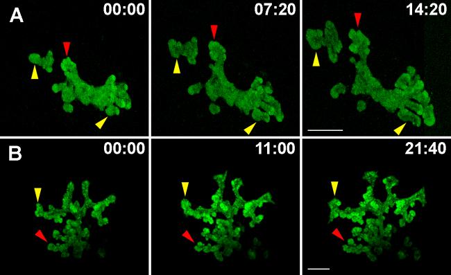
Figure 3. Expansion and branching of the embryonic pancreatic epithelium in real-time.
A. Pancreatic explants were dissected at e10.5 from the Pdx1-GFP transgenic embryos and grown in culture for 4 days before imaging. B. Explants expressing Pdx1-Cre-Z/EG were dissected at e11.5 and grown in culture for 4 days followed by imaging. Still images from time-lapse movies show the overall expansion of the pancreatic epithelium (yellow branches) and branching by budding (red arrows). Time of imaging is shown in hours:minutes. Bar, 200μm.

