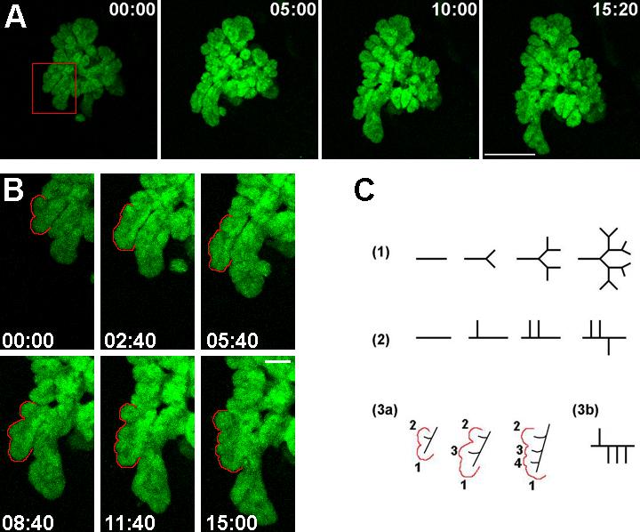
Figure 4. Description of branching in the pancreatic epithelium during morphogenesis.

A. Frames from a time-lapse movie of an e9.5 Pdx1-GFP pancreatic explant grown in culture for 4 days before imaging are shown. Bar, 200μm. B. A magnified view of a region of the growing pancreatic epithelium (indicated in the red box in A) depicts branching in the pancreas. Bar, 50μm. Time of imaging is shown in hours:minutes. Lateral buds appear (B, and C, 3a, labeled 2, 3 and 4) as the “primary” bud (labeled as 1) expands. In C, different modes of branching morphogenesis are represented- (1) type “1-2” terminal bifurcation, (2) type “1-3” lateral budding, and (3) lateral branching observed during pancreas growth. The order of appearance of buds exemplifies this lateral mode of expansion in the pancreas.
