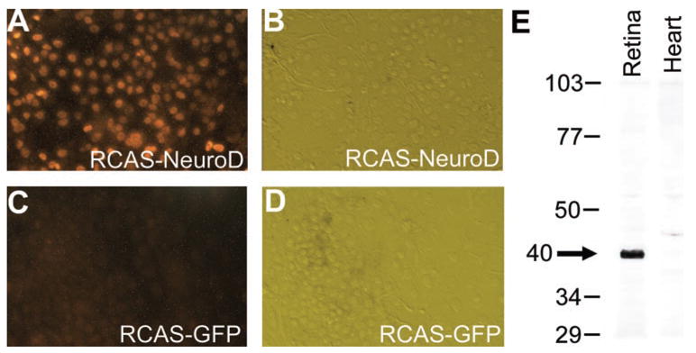
Figure 1.

Immunostaining of cultured, retrovirus-infected RPE cells (A–D) and Western blot of nuclear extracts from the retina and the heart (E) with affinity-purified antibody against NeuroD. (A, C) Fluorescent images of the immunostaining. (C, D) Hoffman images of cells in (A, C). Numbers in (E) indicate molecular mass (in kilodaltons) standards. Arrow: major band present in E9 retina but absent from E9 heart.
