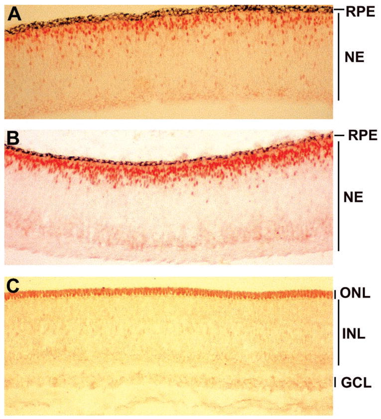
Figure 2.

Expression of neuroD in embryonic chick retina. (A) Anti-NeuroD antibody labeled the nuclei of cells residing at the outer portion of the retinal neuroepithelium at E6. (B) At E7, more cells were labeled with the antibody, and the positive cells appeared to gather at the future position of the ONL. (C) At E9, only the nuclei in the ONL were stained with anti-NeuroD antibody. No nuclei in the INL or the GCL were stained. NE, retinal neuroepithelium; remaining abbreviations are defined in the article text. Magnification: × 50.
