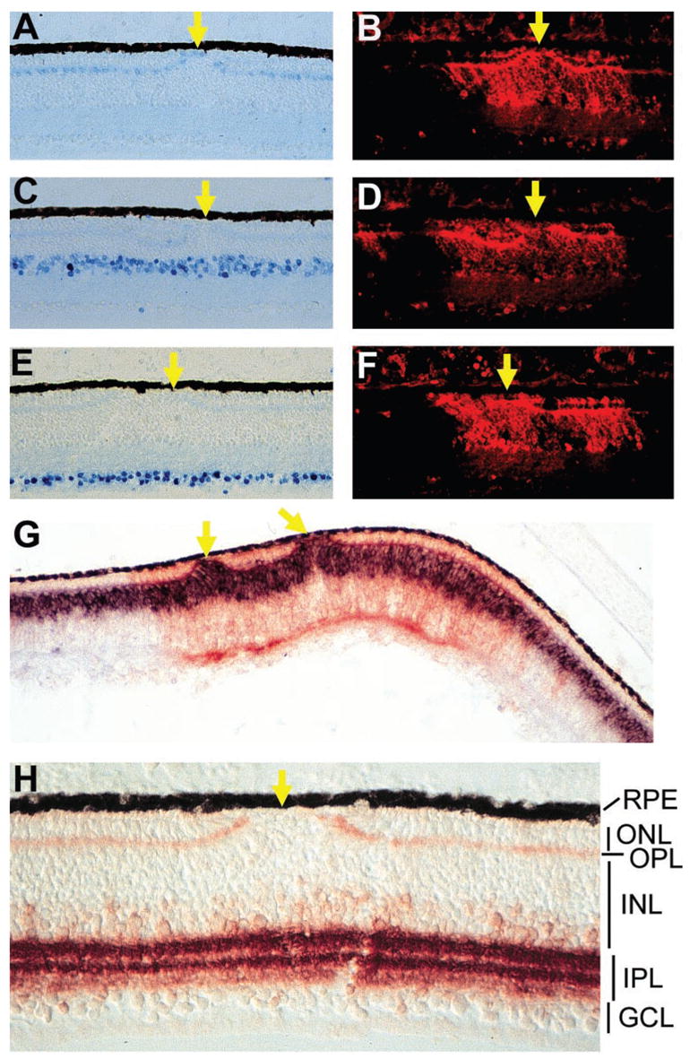
Figure 5.

Nonphotoreceptor neurons in retinas infected with RCAS-En-NeuroDΔC. (A) Horizontal cells at E18 labeled with monoclonal antibody 4F2. (C) Amacrine cells at E18 identified with anti-Ap2 antibody. (E) Ganglion cells at E18 recognized by a monoclonal antibody against Brn3a. (B, D, F) Anti-p27 immunofluorescent staining of the sections in (A), (C), and (E), respectively, to mark regions infected with RCAS-En-NeuroDΔC. (G) Double labeling of an E12 retina for chx10 mRNA (blue) and p27 protein (red). (H) The IPL and the OPL at E18 immunostained with an antibody against SNAP-25. Arrows: places where fewer or no photoreceptor cells are present. Magnification: (A–G) ×50; (H) ×100.
