Figure 1.
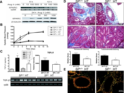
SP−/− mice show attenuated urinary TGF-β1 levels, which correlated with decreases in renal collagen deposition in response to ANG II infusion. A: ANG II treatment results in a concentration-dependent increase in SPARC mRNA expression and protein levels in cultured mouse mesangial cells. mRNA levels were measured by RT-PCR after 6- and 12-hour ANG II treatment at varying concentrations. Protein levels were measured by Western blotting 24 hours after ANG II treatment at varying concentrations. Figures are representative of three independent experiments. B: Tail-cuff measurements of the blood pressures of normotensive (NT) (n = 4) and ANG II-hypertensive (HT) SP+/+ and SP−/− mice (n = 8) in a 14-day period. C: Left, ELISA assay was used to measure urinary levels of active TGF-β1 in the four experimental groups (*P < 0.05 versus SP+/+ NT, #P < 0.05 versus SP−/− NT, and +P < 0.05 versus SP+/+ HT). mRNA expression levels of renal TGF-β1 in hypertensive SP+/+ and SP−/− mice (n = 6 each) as determined by RT-PCR; results shown are representative of three independent experiments (right, quantification of the relative band intensities of TGF-β1 following normalization to corresponding rpS6 internal controls. *P < 0.05 versus SP+/+ HT. Bottom, representative agarose gel of PCR products). D: Determination of collagen deposition in kidneys from SP+/+ and SP−/− HT animals using Masson’s Trichrome staining. Top, perivascular staining of collagen (magnification, ×400). Bottom, tubulointerstitial staining of collagen (magnification, ×400). Graphs indicate the quantification of the Masson’s Trichrome staining by blinded scoring. *P < 0.05 versus SP+/+ HT. E: Picrosirius red staining of renal vessels under polarized light to measure perivascular collagen deposition in SP+/+ and SP−/− HT animals (magnification, ×400). Red color depicts mature collagen fibril staining (SP+/+ HT), and orange-green color indicates the presence of immature fibrils (SP−/− HT).

