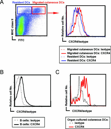
Figure 1.
CXCR4 expression on resident DCs and migrated cutaneous DCs in lymph nodes. A and B: Draining lymph node cells were prepared from mice 24 hours after FITC painting on the abdomen. The profiles show flow cytometric analysis of the cells with the indicated markers. MHC class II+ DCs were subdivided into FITC+-migrated cutaneous DCs and FITC− resident DCs. The profiles show histograms of CXCR4 expression on MHC class II+ FITC+-migrated cutaneous DCs and MHC class II+ FITC− resident DCs (A) and B220+ B cells (B). Data are a representative of three independent experiments. C: Skin organ explants from the ears of the mice were incubated for 24 hours, and the expression of CXCR4 on the emigrated MHC class II+ CD11c+ cutaneous DCs was examined. Data are a representative of three independent experiments. As control, rat IgG2a isotype-matched control was used (A–C).

