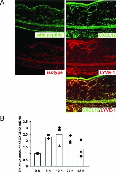
Figure 4.
CXCL12 expression in lymphatic vessels of mouse skin. A: Skin sections from ears of mice treated with DNFB 24 hours prior were stained with goat anti-CXCL12 Ab with or without blocking peptide, and rat anti-LYVE-1 Ab or isotype control Ab, and sequentially immersed with Alexa Fluor 488 rabbit anti-goat IgG and PE-conjugated donkey anti-rat IgG, respectively (the labels are the same color as the reaction product). B: The ears of mice treated with 20 μl of 0.3% DNFB for 6, 12, 24, and 48 hours were isolated. The levels of CXCL12 mRNA were normalized against GAPDH as an endogenous control. The CXCL12 mRNA amounts in the skin from DNFB-treated mice relative to that from non-DNFB-treated mice (0 hours) were induced 6, 12, and 24 hours after hapten application. Filled symbols indicate three independent experiments, and columns represent the average.

