Figure 8.
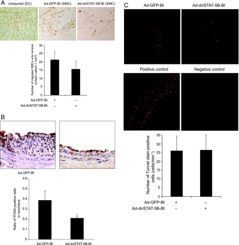
Blockade of STAT-5B signaling suppresses balloon injury-induced SMC migration from media to intima and their neointimal proliferation without affecting cell death. All treatment conditions were the same as in Figure 6 except that 4 days and 1 week after balloon injury rats were sacrificed, arteries were isolated and fixed, and SMC migration from media to intima, SMC neointimal proliferation, and apoptosis were measured as described in Materials and Methods. A: For the measurement of SMC migration, arteries were opened longitudinally, stained for nucleus using anti-histone antibodies (top), and the histone-positive cells in the luminal region were counted (bottom). B: Top panel shows the representative pictures of balloon-injured Ad-GFP- or Ad-dnSTAT-5B-transduced carotid artery sections that were stained for PCNA. The bar graph at the bottom shows the quantitative analysis of the PCNA-positive cells counted in 10 randomly selected fields of the immunostained sections of the balloon-injured Ad-GFP- or Ad-dnSTAT-5B-transduced arteries. C: Top panel shows the representative pictures of balloon-injured Ad-GFP- or Ad-dnSTAT-5B-transduced artery cross sections that were stained with TUNEL kit for detection of apoptosis. The bar graph at the bottom shows the quantitative analysis of the TUNEL-positive cells counted in 12 randomly selected fields of the balloon-injured Ad-GFP- or Ad-dnSTAT-5B-transduced arteries. *P < 0.05 versus Ad-GFP (n = 6).

