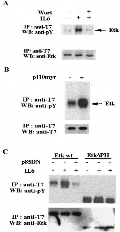
Figure 3.
IL-6 activates Etk through PI3-kinase. (A) Wortmannin (Wort) blocks the activation of Etk by IL-6. The CV-1 cells were transfected with pcDNA3-T7-Etk. At 24 hr posttransfection, the cells were serum starved for 24 hr. The cells were then pretreated with 100 nM wortmannin (+) or dimethyl sulfoxide Mock (−) for 30 min followed by IL-6 treatment for 30 min. The cell extracts were subjected to immunoprecipitation by anti-T7 antibody respectively, followed by Western blot analysis with antiphosphotyrosine antibody (Upper) and anti-T7 antibody (Lower). The tyrosine phosphorylation of the T7-tagged Etk is indicated. (B) A myrislated p110 activates Etk. The CV-1 cells were transfected by pcDNA3-T7-Etk in the presence (+) or absence (−) of pcDNA3-p110 myr. At 24 hr posttransfection, the cells were serum starved for 24 hr. The cell extracts were subjected to immunoprecipitation by anti-T7 antibody respectively, followed by Western blot analysis with anti-phosphotyrosine antibody (Upper) and anti-Etk antibody (Lower). (C) A dominant negative p85 blocks the activation of Etk by IL-6. The CV-1 cells were transfected with pcDNA3-T7-Etk (wild type) or pcDNA3-T7-EtkΔPH(deletion of PH domain) in the presence (+) or absence (−) of pcDNA3-p85DN. At 24 hr posttransfection, the cells were serum starved for 24 hr followed by IL-6 treatment for 30 min. The cell extracts from each treatment were subjected to immunoprecipitation by anti-T7 antibody, followed by Western blot analysis with antiphosphotyrosine antibody (Upper) and anti-Etk antibody (Lower).

