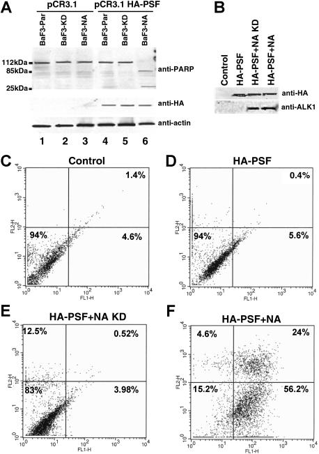
Figure 7.
PSF overexpression induces apoptosis in NPM/ALK-expressing cell lines. (A) BaF3-Par, BaF3-KD, and Baf3-NA cells were electroporated with pCR3.1 plasmid alone (lanes 1-3) and with pCR3.1 HA-PSF (lanes 4-6). After 30 hours of culture, the cell extracts from 5 × 10⁶ cells were lysed and subjected to denaturing SDS-PAGE and Western blotting with an anti-PARP antibody. PSF expression and protein loading were controlled by anti-HA and antiactin Western blotting. (B-F) 293T cells were transiently transfected with pCR3.1 empty vector (CON) or HA-PSF alone or together with kinase-dead or wild-type NPM/ALK. The efficiency of transfection was determined by anti-HA and anti-ALK1 Western blotting at 72 hours after transfection (B). Apoptosis was assessed at the same time point by Annexin V analysis in control cells (C), cells transfected with HA-PSF alone (D), or together with kinase-dead NPM/ALK (E) (HA-PSF + N/A-KD), or wild-type NPM/ALK (F) (HA-PSF + NA). C-F, live cell percentages are shown in the bottom left panels; apoptotic cell percentages in the bottom right panels, and dead cell percentages in the top right panels.

