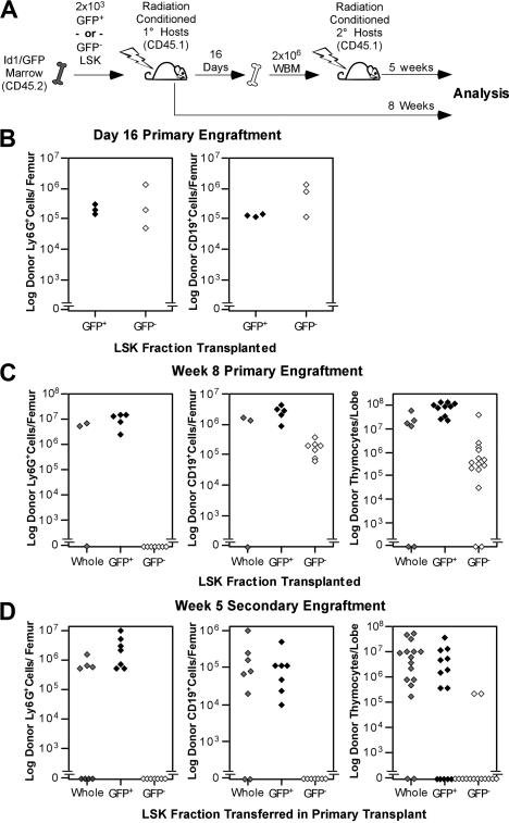
Figure 3.
Functional evaluation of Id1/GFP+LSK. (A) Schematic representation of the transplantation protocol used to determine the presence of LT-HSCs in Id1/GFP-expressing LSK cells. Subsets were sorted from CD45.2 Id1GFP/GFP donors. GFP+ preparations transplanted were at least 10-fold enriched for Id1/GFP+ cells compared with unseparated LSK cells. GFP− preparations included no more than 0.8% GFP+ cells. Primary and secondary hosts were treated prior to transplantation with sublethal doses of radiation. Separate cohorts of CD45.1 host mice each received 2 × 10³ sorted Id1/GFP+, Id1/GFP−, or whole LSK cells. After 16 days, WBM was collected from a subset of primary hosts. Marrow was pooled from hosts receiving the same LSK subsets. A total of 2 × 10⁶ cells were then transferred to each mouse in fresh cohorts of CD45.1 hosts. The remaining primary hosts were evaluated for marrow and thymic engraftment 8 weeks following transplantation. Secondary hosts were assayed for engraftment 5 weeks after secondary transplantatoin. (B) Day-16 analyses of primary engraftment. One femur was saved from each mouse for analysis, while marrow from the remaining femur and both tibia was used for secondary transplantations. For analysis, marrow was immunofluorescently labeled with antibodies against the donor CD45 isoform, Ly6G, and CD19. Donor fractions observed from flow cytometry were multiplied by cell counts to determine total donor-derived cells per femur in either the B-cell or myeloid lineages. (C) Long-term primary engraftment from Id1/GFP LSK subsets. Primary hosts were evaluated for engraftment 8 weeks after transplantation. Bone marrow was analyzed as described in panel B. In addition, thymic lobes from each host were evaluated separately for donor progeny. (D) Secondary engraftment analyses. Marrow and thymus from secondary hosts were analyzed as in panels B and C 5 weeks following secondary marrow transfer. For all panels, progeny from unseparated (whole) LSK, Id1/GFP+LSK, and Id1/GFP−LSK cells are represented by gray, black, and white symbols, respectively. Each symbol represents the total donor-derived progeny of the indicated lineage for 1 femur or thymic lobe, shown in log scale.

