
Figure 2.
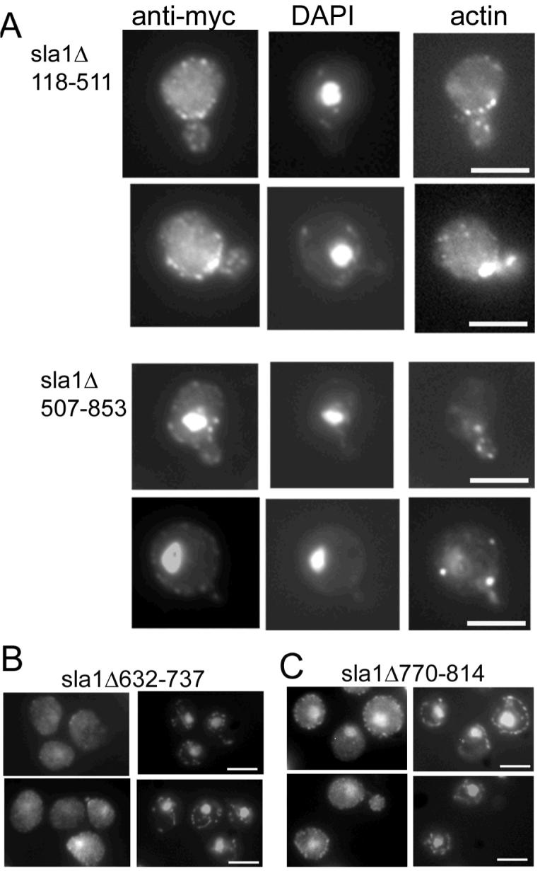
A. Cells expressing mutant forms of Sla1p (Δ118-511, or Δ507-853) were grown to log phase and then fixed and processed for immunofluorescence and staining with rhodamine-phalloidin. Co-staining was achieved by mounting cells in anti-fade solution containing the DNA dye DAPI. In the upper panels Sla1Δ118-511p can be seen to exhibit only staining to punctate cortical sites while in the lower set of panels, cells expressing Sla1Δ507-853 can be seen to show a high level of localisation to the nucleus. B. Strains carrying mutant Sla1 proteins with deletions of either nuclear export signal were generated and cells were grown, then fixed and processed for immunofluorescence. Of the mutants Sla1Δ623-737 gave a low level of nuclear stain similar to wild-type, while the nuclear staining for Sla1Δ770-814 was more marked and resembled the larger deletion shown in panel A. Bars = 5 μm