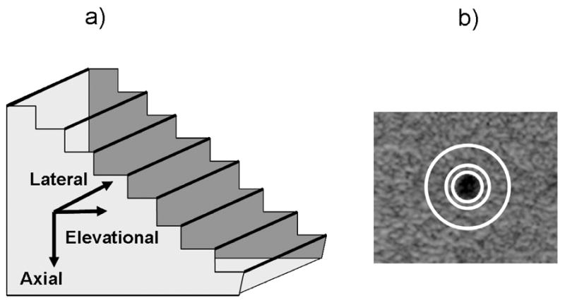
Fig. 2.

a) Step-shaped string test object with 25 μm silver wires acting as line targets to assess the LSF of the system in all three directions. b) Anechoic cylinder within contrast test object for determining contrast with and without imaging through a mammographic paddle. Mean cylinder amplitude was calculated within the innermost white circle and mean background amplitude was calculated in the area between the two outermost white circles. Mean signal strength for each paddle and no paddle was also calculated from a uniform speckle region within this phantom over a 4 cm depth.
