Figure 2.
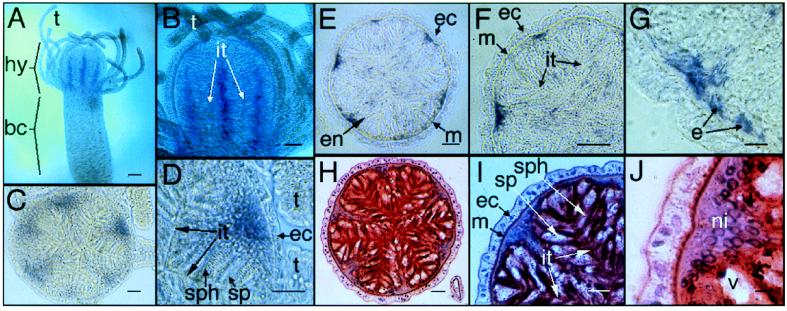
In situ hybridization of Cn-ems. (A and B) Whole mounts. (A) Longitudinal stripes (purple/blue) representing Cn-ems expression are restricted to the hypostome and are absent from the body-column. (B) Stripes are localized to the center of taeniolae. (C and D) Thick sections. (C) Cross-section of the hypostome area, showing endodermal expression of Cn-ems in the mid-line of taeniolae bases. (D) Close-up of C, showing details of taeniolae. (E–J) Thin sections. (E–G) Immunostaining showing localization of Cn-ems mRNA. (H–J) Hematoxylin and eosin staining. bc, body-column; e, endodermal epithelial cell; ec, ectoderm; en, endoderm; hy, hypostome; it, inter-taeniolae border; m, mesoglea; ni, nuclei; sp, spumeous cell; sph, spherulous cell; t, tentacle; v, vesicle of a spumeous cell. [Magnifications: ×40 (A), ×100 (B), ×200 (C, E, and H), ×400 (D, F, and I), and ×1,000 (G and J); bars = 100 μm (A), 50 μm (B), 20 μm (C–F, H, and I), 5 μm (G and J).]

