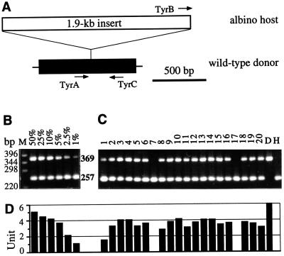
Figure 2.
PCR detection of Mes1-derived chimeras. (A) Schematic structure of the tyrosinase gene in the wild-type Mes1 donor (HB32C) and albino host strains (i1). Only the first exon (black box) of the gene and the 1.9-kb insert (open box) interrupting the exon are shown. PCR primers are represented by arrows. TyrA and TyrC define a fragment of 369 bp specific to the donor strain, whereas TyrB and TyrC give rise to a 257-bp fragment unique to the host strain. (B) Sensitivity of the PCR assay. Lane M, 1-kb marker (GIBCO), with sizes shown in base pairs. Numbers in percentages indicate proportions of donor strain-derived DNA diluted with that of the host strain. The 369-bp, donor-specific band is detectable if the donor DNA represents at least 1% of the input DNA. (C) Screening of Mes1-injected embryos from the third transplantation experiment (Table 1). The 369-bp, donor-specific band is evident in 18 of 20 hatchlings and fry from Mes1-transplanted embryos (lanes 1–20) and in the donor (lane D) but not the host (lane H) strain. All host embryos display the 257-bp band. Lanes 1–20, pigmented (lanes 1 and 2) and nonpigmented (lanes 3–20) fry (lanes 1–11) and hatchlings (lanes 12–20). The second pigmented chimeric fry (lane 2) is shown in Fig. 1E. (D) Graphs of intensities of the 369-bp, donor-specific band in the samples shown in B and C. The intensity of the 369-bp band in the 1% lane (B) is equivalent to 1 arbitrary unit.

