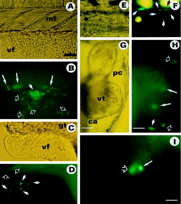
Figure 4.
Chimeric fry from Mes1 cells transiently expressing GFP. (A, C, E, and G) Bright-field micrographs. (B, D, F, H, and I) Dark-field fluorescent micrographs. (A and B) GFP-expressing Mes1 cells in the myotome (mt; large arrows) and the ventral fin (vt) as clustered (small arrow) and single (hollow arrows) cells. (C and D) GFP-expressing Mes1 cells in the ventral fin (vf; arrows) and the gut (gt; hollow arrows). Note the distinct epithelial phenotype of GFP-expressing cells that are morphologically indistinguishable from surrounding recipient cells. The gut is recognizable by its background autofluorescence. (E and F) GFP-expressing Mes1 cells in the trunk (arrows). GFP-positive cells show green fluorescence and can be distinguished from large, yellow autofluorescent recipient pigment cells. (G and H) GFP-expressing Mes1 cells in the ventral body wall surrounding the pericard (pc; hollow arrows), the ventricle (vt; large arrows), and conus arteriosus (ca; small arrow) of the heart. (I) GFP-expressing Mes1 cells in the atrium (solid arrow) and ventricle (hollow arrow) of the heart. The anterior is to the right, the dorsal side is up. [Bar = 50 μm (A–F and I) and 10 μm (G and H).]

