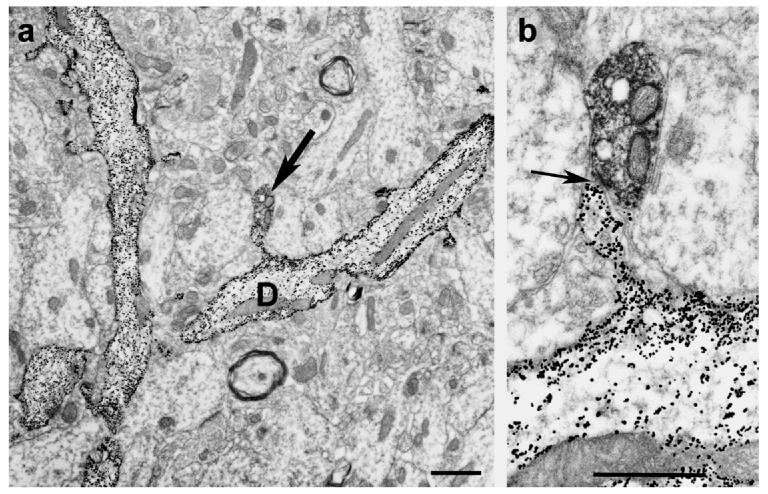
Fig. 1.
Low (Panel a) and high power (Panel b) electron micrographs (received from Dr. Michael Frotscher) show the result of a combined Golgi impregnation and ChAT immunostaining experiment. On Panel a, Golgi-impregnated (gold-toned) spiny granule cell dendrites are seen. Arrow on the same panel points at a ChAT immunoreactive bouton contacting the spine head of the dendrite (D). Panel b shows the asymmetric synaptic contact (arrow) between the two profiles.
Bar scales= 1 μm.

