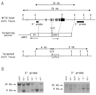
Figure 1.
Mutation of the Usf1 locus. (A) Targeting strategy. (Top) Structure of the wild-type Usf1 gene with solid boxes representing exons. The sizes of the restriction fragments detected by the indicated probes in wild-type DNA are shown above. E, EcoRI; V, EcoRV. (Middle) The gene-targeting vector. Open boxes, Usf1 homologous regions; neo, the PGK-neo expression cassette that introduces novel EcoRI and EcoRV restriction sites; tk, the MC1-tk expression cassette used for negative selection. The arrows beneath neo and tk indicate the direction of transcription of each cassette. (Bottom) Structure of the targeted locus, with the sizes of the restriction fragments detected by the Southern probes shown above. (B) Southern blot showing genotypes of newborn mice from a heterozygote mating. EcoRI-digested tail DNA was hybridized with the 3′ probe, and EcoRV-digested tail DNA was hybridized with the 5′ probe. The wild-type and mutant bands are shown in each case. Lanes: +/+, wild type; +/−, heterozygous mutant; −/−, homozygous mutant.

