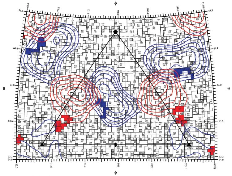
Figure 7.
A roadmap showing the location of surface residues of CPV-2 that affect the binding of the different antibodies to the capsid structures. A single asymmetric unit of the capsid is shown. Two antigenic sites have been previously defined, and designated as (A) and (B) through the analysis of mutations that affected antibody binding or by cross-competition. Residues that affect antibody binding to some A-site antibodies are shown in red, and those affecting B-site antibodies are shown in blue. CryoEM density corresponding to Fab14 as bound to the A site and Fab15 as it interacts with the B site are projected to the surface of the virion and indicated as red (Fab14) or blue (Fab15). The projection of Fab density obtained from virus-Fab complex shows the A and B sites as previously described, but with some possible overlap between the antibodies binding to the two sites (Hafenstein et al., 2006).

