
Fig. 1.
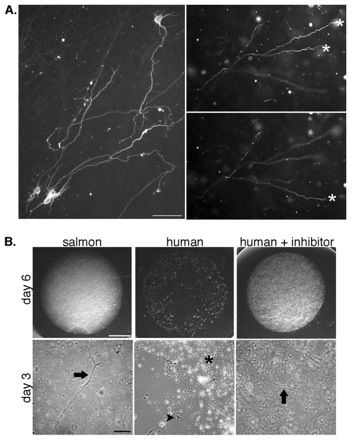
Neurons embedded in fibrin extend neurites in three dimensions and rapidly degrade mammalian fibrin. A, Neurons were fixed and immunostained with the neurite marker TuJ1 (which labels both axons and dendrites in embryonic neurons) after growth in gels for 6 days (left panel) or 2 days (right panels). Images of neurites at multiple focal planes were combined in a montage to show the extent of neurite growth (left panel). Right panels show a region of a fibrin gel imaged at two focal planes to demonstrate that neurites have extended in the z-plane. Asterisks mark neurites that are in focus in each plane. Scale bar is 50 μm. B, Imaging of the entire gel shows that human gels without plasmin inhibitor are entirely degraded by 6 days (upper panels, scale bar is 200 μm). Higher magnification images of gels at an earlier time point (3 days, lower panels, scale bar is 100 μm) show areas of degradation of the human gels that have left the cells on the coverslip surface (arrowhead, middle panel) next to the residual gel (asterisk). Neurons remain embedded in the salmon fibrin and human fibrin with plasmin inhibitor (arrows).