
Figure 1.
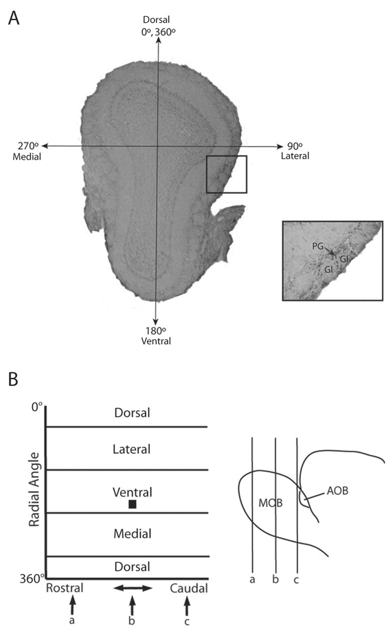
Identification and mapping of activated glomeruli in the main olfactory bulb in a WT female mouse following exposure to E female volatile urinary odors. (A) An activated glomerulus (Gl) was defined as having two 90° arcs or a 180° continuous arc of Fos-IR in the periglomerular cells (PG) surrounding the glomerulus. The representative coronal section shown in panel A is located at point b in panel B. A central axis was established (0–180°) using anatomical landmarks, extending from the dorsal mitral cell layer to the ventral mitral cell layer. The lateral, ventral, medial, and dorsal portions of the bulb are located at radial angles of 90°, 180°, 270°, and 360° respectively. The location of each activated glomerulus was recorded using the rostral-caudal distance through the bulb as well as the radial angle, and a composite map was formed showing areas of greatest activation throughout the bulb. In panel B, the location of the slice (b) as well as the activated glomeruli highlighted in the inset are depicted by the black box located in the lateral region of the 2-dimensional map, ~120° from the central axis.