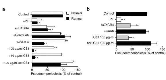
Figure 8.
Inhibition of B-cell pseudoemperipolesis of RA FLSs by CXCR4 and VLA-4 antagonists. (a) Nalm-6 (open bars) or Ramos cells (filled bars) were preincubated with pertussis toxin (PT), αCXCR4 mAb’s, control mAb’s, αVLA-4 mAb’s, 100 μg/ml or 10 μg/ml of CS1 peptide, or a control peptide, as indicated on the left-hand side. Cells then were incubated on RA FLSs and allowed to migrate beneath the FLSs for 2 hours. Then, the FLS layer containing the migrated cells was harvested, and the relative numbers of migrated cells were determined by flow cytometry. The bars represent the mean (± SD) B-cell migration relative to that of untreated samples. AThe difference between the percent migration under a given condition is significantly less than that noted for same cell population for FLSs in the absence of inhibitors (e.g., P values < 0.05, Bonferroni’s t test). (b) Pseudoemperipolesis of normal blood B cells beneath RA FLSs was also inhibited by PT, αCXCR4 mAb’s, or CS1 peptide. The bars represent the mean (± SD) relative B-cell migration of B cells from five different donors. ASignificant inhibition of migration with P values < 0.05 using Bonferroni’s t test.

