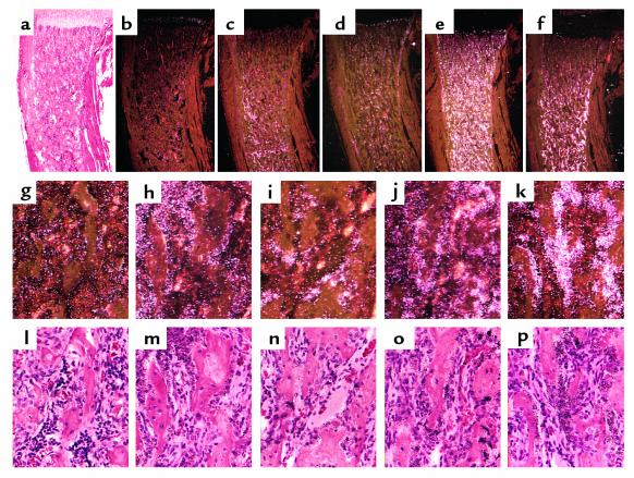
Figure 3.
In situ hybridization analysis of trabecular bone. In situ hybridization with the 35S-labeled alkaline phosphatase (a, b, g, l), collagen I (c, h, m), collagenase 3 (d, i, n), osteopontin (e, j, o), and osteocalcin (f, k, p) cRNAs in serial sections of decalcified proximal tibia from 2-week-old CL2 mouse. Higher-magnification images of the trabecular area are shown (g–p). The sections were counterstained with hematoxylin and eosin; bright-field (a, l–p) and dark-field (b–k) views are shown.

