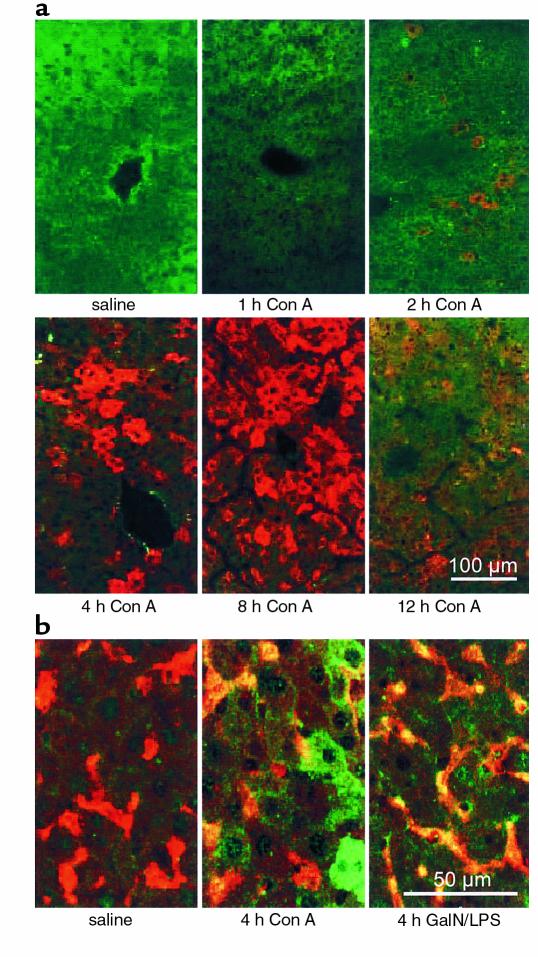
Figure 2.
Detection of iNOS in livers of Con A–treated mice by immunofluorescence staining. (a) Con A–induced iNOS expression in livers of BALB/c mice was detected by immunofluorescence staining of liver cryostat sections (rabbit anti-mouse iNOS antiserum, secondary goat anti-rabbit IgG tagged with Cy3; red fluorescence). (b) Costaining of iNOS (rabbit anti-mouse iNOS antiserum, secondary swine anti-rabbit IgG tagged with FITC; green fluorescence) and Kupffer cells (BM8, secondary goat anti-rat IgG tagged with Texas red; red fluorescence) 4 hours after Con A or GalN/LPS treatment. All sections were examined by confocal laser-scanning microscopy. Costaining is represented by yellow fluorescence. Seen in a and b is one example of three independent experiments, respectively.

