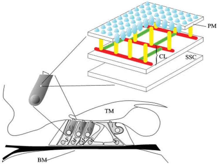
Fig. 1.

Diagram of the organ of Corti depicting OHC, IHC and support cell location. The OHC lateral wall consists of the plasma membrane (PM), cortical lattice (CL), and subsurface cisternae (SSC). The motor protein prestin (blue) is highly enriched in the PM. The cortical lattice, consisting of F-actin (red) and spectrin filaments (green), is connected to the plasma PM by pillar-like proteins of unknown identity (yellow). Two layers of SSC are depicted below the cortical lattice. O, outer hair cell; I, inner hair cell; S, support cell; BM, basilar membrane; TM, tectoral membrane. [Color figure can be viewed in the online issue, which is available at www.interscience.wiley.com.]
