Fig. 3.

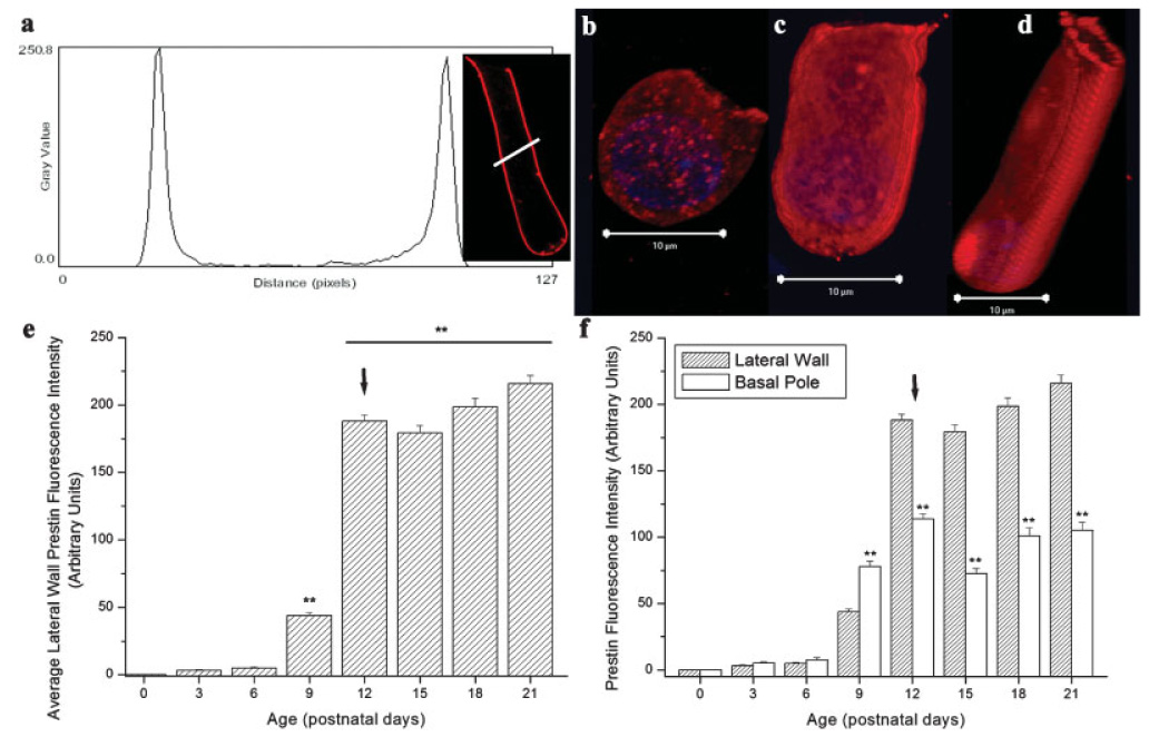
Lateral wall prestin accumulation in developing OHCs. (a) Representative prestin fluorescence intensity profile (P21). (b–d) 3-D reconstructions of P6, P9, and P12 prestin-labeled isolated OHCs, respectively. (e) Significant differences in lateral wall prestin accumulation, measured by alterations in fluorescence intensity, were observed in developing OHCs (F(7,571) = 640.1, P < 0.01). Lateral wall prestin accumulation was greatest immediately before hearing onset (P9–P12). After hearing onset (P12), the OHC lateral wall contained significantly more prestin than before hearing onset (P9). (f) Comparison of average lateral wall and basal pole prestin labeling during postnatal development. At and after the onset of hearing, prestin was significantly reduced in the basal pole. Arrow = hearing onset, ** = P < 0.01.
