Figure 3.
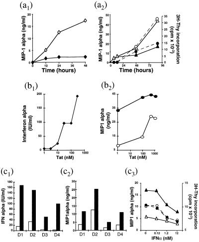
Effect of IFNα and Tat on the regulation of C-C chemokines production. (a) Effect of IFNα on immune cells. (a1) Kinetics of MIP-1α production by PBMC from a seronegative donor, cultured in the presence of sheep anti-IFNα (—⧫—) or control serum (—⋄—) at 1:200 dilution. (a2) Kinetics of T cell proliferation (—○— —•—) and of MIP-1α (—▵— —▴—) production in PBMC from a representative donor, cultured in the absence (—○— —▵—) or the presence (—•— —▴—) of recombinant IFNα at 10 nM. Each experiment was repeated at least three times. MIP-1α levels were measured by ELISA; cell proliferation by 3H-thymidine incorporation and results were expressed as cpm. (b) Effect of HIV-1 Tat on macrophages. (b1 and b2) Monolayers of purified macrophages were pretreated for 3 h at 37°C in HL-1 medium with varying concentrations of Tat protein. Tat pretreated cells were then cultured in the presence of PHA-P for 24 h. (b1) SN were collected for IFNα titration (41). (b2) Cells were cultured in the presence (—•—) or the absence (—○—) of IFNα (12.5 nM), and SN was collected after 24 h for MIP-1α titration. (c) Effect of Tat on PBMC. PBMC from seronegative donors were pretreated for 3 h with (▪) or without (□) Tat (concentration 200 nM) before activation with anti-CD3 Abs and cultured for 3 days in the presence of IL-2. SN were tested in c1 for IFNα and in c2 for MIP-1α production. (c3) SN of cells pretreated with (—•— —▴—) or without (—○— —▵—) Tat protein were tested for MIP-1α production (—▵— —▴—) and for T cell proliferation (—○— —•—). D1, D2, D3, and D4 = donors 1, 2, 3, and 4.

